How Is Jock Itch Treated?
Otherwise known as Tinea Cruris, it can have a similar presentation and symptoms as other rashes.
Tinea cruris is a common problem that mainly affects male adolescents and adults. This fungal infection, also known as jock itch—and sometimes Dhobi itch or eczema marginatum1—must be treated, as it will not go away on its own. Tinea cruris is widespread globally, particularly in places with high humidity and temperatures, such as the tropics.1,2Other risk factors include constant skin-on-skin friction, diabetes, excess sweating, obesity, and poor hygiene.1,3,4The condition is on the rise among women, especially those who wear clothing or stockings that are too tight; it is rarely found in children.5,6
Tinea infections are often caused by dermatophytes. The dermatophyte most implicated in tinea cruris is Trichophyton rubrimus, which is the same causative agent for tinea pedis. Other contributing species include Epidermophyton floccosum, Microsporum canis, and Trichophytom mentagrophytes1,6,7; occasionally a secondary infection from bacteria or yeast is present.7
Self-inoculation to the groin region may occur from tinea pedis on the foot, as the 2 infections are commonly associated with each other.1,7Another possibility of infection is indirect contact through sharing sports equipment or towels.1
CLINICAL PRESENTATION
Initial symptoms of tinea cruris consist of a unilateral erythematous rash starting in the crease of the groin that can spread onto the upper thigh.1,3The rash typically forms as a half-moon shape, with distinct squamous borders and scaling. Sometimes, vesicles progress out of the crease of the groin.
The rash may spread to the other thigh (if it is not initially infected), gluteal folds, and the pubic, suprapubic, and perianal regions, usually sparing the penis and scrotum. Lesions often have a tendency for central clearing, and satellite sites are infrequent. Itching is a common complaint.1,5-9Pain described as burning and stinging may also be present, especially in cases with a secondary infection.4
DIFFERENTIAL DIAGNOSIS/WORKUP
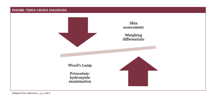
Tinea cruris needs to be distinguished from other rashes (figure1,3,5,6). Possible differential diagnoses includes candidiasis, erythrasma, intertrigo, pityriasis versicolor, and psoriasis. Candidiasis and intertrigo also cause groin rashes. Candidiasis has a characteristic “beefy red” appearance, with intense erythema, non—well-defined borders, andwhite pustules. Numerous small satellite sites are also common. Intertrigo is similar to tinea cruris, as it has a half-moon–shaped rash in the groin region, which can spread onto the thigh from moisture accumulation.4-6Pityriasis versicolor can sometimes be present in the groin area; however, asymptomatic, noninflamed lesions present in this rash can aid in distinguishing the 2 conditions.
Additional considerations are contact dermatitis and erythrasma. A Wood’s lamp can help differentiate tinea from other skin infections, as under it, erythrasma appears coral red and fungal infections often fluoresce as a light green. The location of lesions on other body parts may help identify other rashes, such as dermatitis or psoriasis.1,3Accurate diagnosis requires a sample acquired from the progressing scale borders for a potassium-hydroxide exam.5

TREATMENT
Tinea cruris needs to be treated. Some general suggestions for what can be done prior to the need for prescription medication include the following:
- Bathing in nonsoapy water, until skin clears, as infected skin can be irritated by soap
- Drying oneself well after exercising or swimming and not changing into wet clothing afterward
- Using topical OTC medications, such as ointments, powders, and sprays4
- Wearing well-fitted clothing that is not too tight
Topical medication is efficacious for most patients with tinea cruris, and these include allylamines, butenafine, ciclopirox, imidazole, or tolnaftate. Treatment should be applied to the affected areas twice daily for at least 10 to 14 days, sometimes up to 3 weeks, depending on the extent of the rash. Completion of treatment is essential.1,2,5Oral medication should not be used unless topical treatment fails.10Symptoms generally improve within 2 to 3 weeks of treatment, but recurrence frequently occurs. Complications are rare, but allergic reaction or a secondary bacterial infection may appear.4,10
CONCLUSION
As tinea cruris can have a similar presentation and signs/symptoms of other conditions, a careful examination and using appropriate tools for diagnosis is essential. Steps can be taken to reduce its development and spread, but intervention is crucial, as the infection will not resolve by itself.
REFERENCES
- Metin A, Dilek N, Demirseven DD. Fungal infections of the folds (intertriginious areas).Clin Dermatol.2015;33(4):437-447. doi: 10.1016/j.clindermatol. 2015.04.005.
- Sahoo AK, Mahajan R. Management of tinea corporis, tinea cruris, and tinea pedis: a comprehensive review.Indian Dermatol Online J. 2016;7(2):77-86. doi: 10.4103/2229-5178.178099.
- Clebak KT, Malone MA. Skin infections.Prim Care. 2018;45(3): 433-454.doi:10.1016/j.pop.2018.05.004.
- Safran MR, Zachazewski J, Stone DA. Tinea cruris (jock itch). In: Safran MR,Zachazewski J, Stone DA, eds. Instructions for Sports Medicine Patients. 2nd ed. Philadephia, PA: Elsevier; 2012.
- Kaushik N, Pujalte CG, Reese ST. Superficial fungal infections.Prim Care.2015;42(4):501-516. doi: 10.1016/j.pop.2015.08.004.
- Panthagani AP, Tidman MJ. Diagnosis directs treatment in fungal infections of the skin.Practitioner.2015;259(1786):25-29, 3.
- Dawson AL, Dellavalle RP, Elston DM. Infectious skin diseases: a review and needs assessment.Dermatol Clin.2012;30(1):141-151. doi: 10.1016/j.det.2011.08.003.
- Hay R. Superficial fungal infections. Medicine Journal UK. 2017;45(11):707-710.doi: 10.1016/j.mpmed.2017.08.006.
- El-Gohary M, van Zuuren EJ, Fedorowicz Z, et al. Topical antifungal treatments for tinea cruris and tinea corporis. Cochran Database Syst Rev. 2014;8:CD009992 doi:10.1002/14651858.CD009992.pub2.
- Decker CF. Skin and soft tissue infections in the athlete.Dis Mon. 2010;56(7):414-421. doi: 10.1016/j.disamonth.2010.05.002.

